Digital dental X-rays: clearer images, lower dose
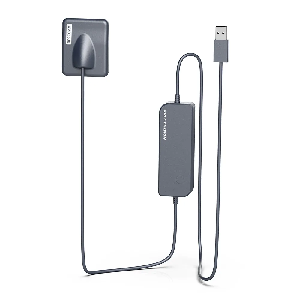
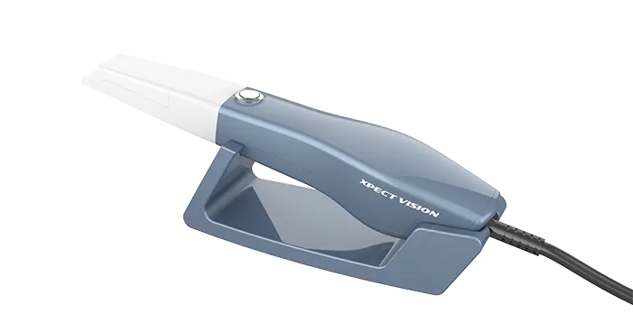
Digital sensors (RVG) changed intraoral imaging: the image is on screen within seconds, you can zoom, adjust contrast and measure canal length right away, usually with a lower radiation dose than conventional film.
Tips for the best image
- Right exposure time: a digital sensor needs less time than film, and a longer exposure does not make a clearer image, only a higher dose. Start from the suggested setting for each tooth and adjust it to the patient's size.
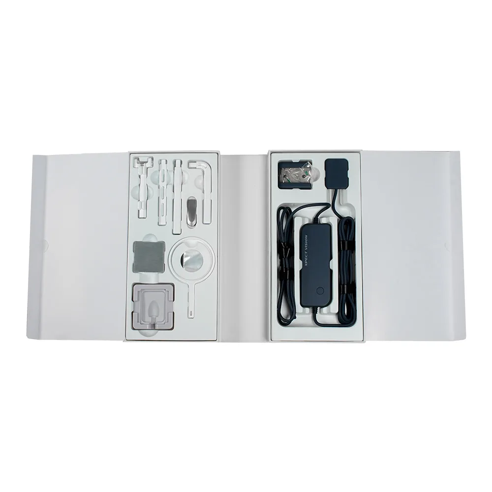
- Sensor holder: positioning holders with the paralleling technique reduce distortion and make images comparable from one visit to the next.
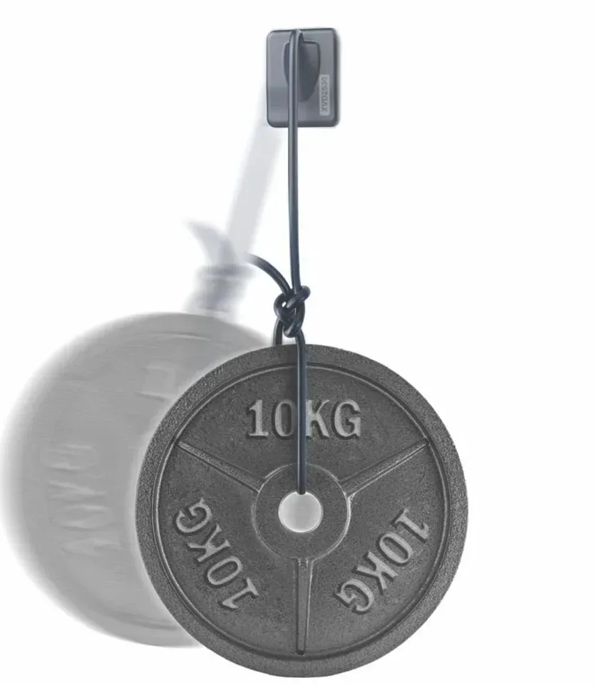
- Protect the sensor: use a new barrier sleeve for every patient, and never bend the cable or let the patient bite on it; it is the part most likely to break.
- Lower the dose: a rectangular collimator shrinks the exposed area and cuts the dose noticeably; always follow approved radiation-protection guidelines.
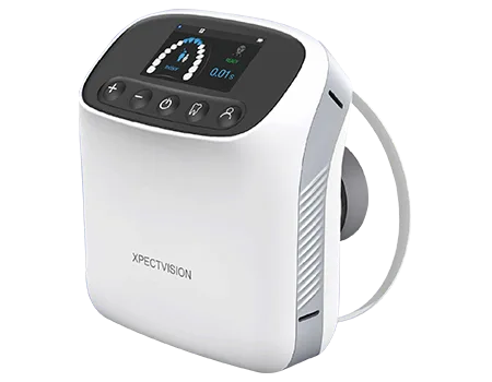
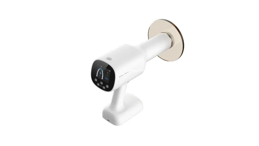
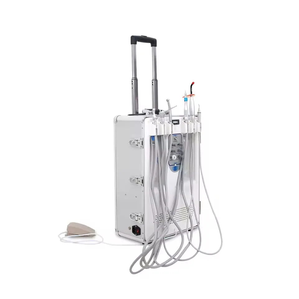
With portable X-ray units, imaging is now possible right at the chair without a separate room, as long as the manufacturer's instructions and radiation-protection standards are followed.